What Actually Happens During a Breast Ultrasound
August 4, 2026
Why Your Doctor May Recommend Breast Ultrasound
A breast ultrasound creates detailed pictures of breast tissue using high-frequency sound waves. Doctors often recommend it after finding a lump, unusual pain, nipple discharge, or an area needing a closer look on a mammogram. It can also help show if a mass is fluid-filled or solid.
If you’re scheduled for a breast ultrasound, knowing the process can help you feel less uncertain. The exam is noninvasive, doesn’t use ionizing radiation, and requires little preparation.
A Quick Look at the Numbers
Breast cancer remains common in the United States. The American Cancer Society estimates 321,910 new invasive cases among women in 2026. It also estimates 60,730 new DCIS cases, while breast cancer is expected to cause about 42,140 deaths. These figures show why a new breast concern deserves prompt evaluation.
What to Do Before Your Appointment
Most breast ultrasound exams require little preparation. Wear comfortable clothing that’s easy to remove from the waist up. You’ll usually change into a gown.
Bring imaging history if requested. Tell the team about symptoms, prior procedures, or recent mammograms.
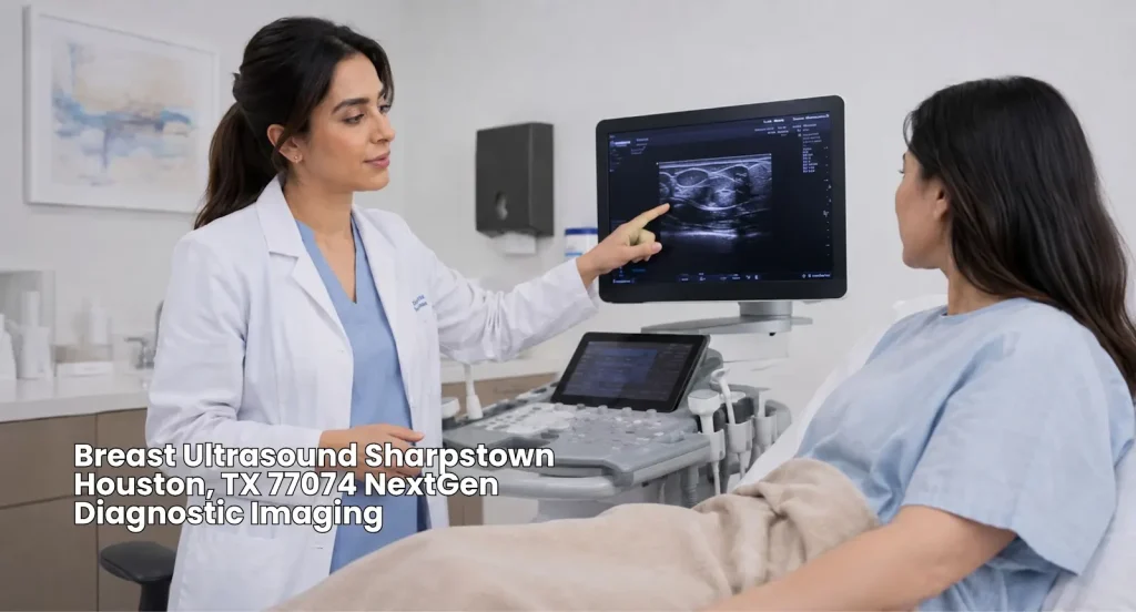
What Happens When You Arrive at NextGen Diagnostic Imaging
At NextGen Diagnostic Imaging, expect a straightforward check-in. Staff may confirm your appointment, symptoms, and reason for the exam.
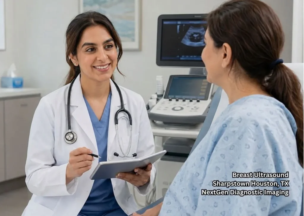
You’ll then meet the sonographer, who may ask you to point out a lump or tender area. This helps guide the examination.
What Happens During the Scan
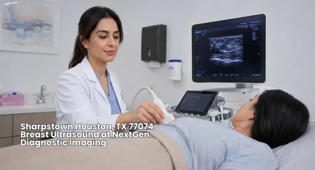
You’ll lie on your back, sometimes with an arm raised above your head. The sonographer applies clear gel to the skin. A handheld device called a transducer then moves across the breast.
The transducer sends sound waves into tissue and captures returning signals. A computer turns them into live images. The sonographer may take several views of an area that looks different.
Is a Breast Ultrasound Painful?
Most people find the test comfortable. You may feel pressure over a tender area, but the exam is generally not painful. Radiologist Dr. Elissa Price calls it “a safe, noninvasive test.”
What the Sonographer Is Looking For
The scan can characterize a breast abnormality. A key question is if an area appears cystic, solid, or mixed. Ultrasound can also show tissue in real time and, in selected cases, blood flow.
A finding on ultrasound doesn’t automatically mean cancer. Many abnormalities are benign. Once the images are reviewed, the radiologist documents the findings in a report for your doctor.
Why Mammograms and Ultrasound Can Work Together
Ultrasound isn’t a replacement for mammography. Mammograms remain important for screening, while ultrasound can provide more information about specific findings, including dense tissue or a lump.
If you’re looking specifically for a breast ultrasound in Sharpstown, ask your doctor why the exam was recommended and how it fits with your other breast imaging.
What Happens After the Examination
After examining the images, the radiologist provides the findings in a report for your doctor. Depending on the findings, you may need routine screening, follow-up imaging, or a biopsy.
A BI-RADS assessment may appear in your report. BI-RADS 2 indicates a benign finding, while BI-RADS 3 generally calls for short-term follow-up. Your physician can explain your specific result.
Finding Your Way Around Sharpstown
Sharpstown is a busy Houston community with several major roads and nearby services. Checking your route before leaving can help make your visit easier.
If you’re using Houston Christian University as a nearby landmark, include it when planning your route. Check your map app for route options before heading to the location.
A Useful Route Reference
If you’re traveling near Sharpview Dr & Cannock Rd, check current traffic before leaving. This crossing can serve as a helpful reference point when navigating the area.
For another reference, enter 29.698716762129813, -95.50933967480091 into your preferred map app to locate the area more precisely.
When a Lump Needs More Explanation
A new breast lump can be unsettling, but getting the right imaging can help provide clearer answers. Our guide on breast ultrasound after finding a lump explains why your doctor may recommend an ultrasound and what to expect next.
Get Directions from Sharpstown to NextGen Diagnostic Imaging
Take the Next Step With Confidence
A breast ultrasound is a simple, noninvasive exam that can provide useful information about breast tissue and help guide your next step in care. If your doctor recommends imaging, bring your questions, follow your healthcare team’s guidance, and book now with NextGen Diagnostic Imaging.
FAQs
1. How long does a breast ultrasound take?
Scan time varies by the exam and images needed. Additional views can make it longer.
2. Can I eat before a breast ultrasound?
Yes. Breast ultrasound typically doesn’t require fasting.
3. Does breast ultrasound use radiation?
No. Ultrasound uses sound waves, not ionizing radiation.
4. Can ultrasound tell if a lump is cancer?
Ultrasound can describe a lump and distinguish cystic from solid findings. A biopsy may be recommended when imaging shows a suspicious abnormality.
5. Will I get my results immediately?
Not always. A radiologist will analyze your images and deliver a professional interpretation report. Your healthcare provider will typically share the results with you once the report is available.

NextGen Scans
Be part of a team driven by compassion and a commitment to putting patients first. At NextGen Scans, the care you provide has a meaningful impact every single day.
or Call us at
(346) 487-7037
Think You Feel Fine? These 3 Findings Often Surprise Ultrasound Patients
Feeling healthy is a good sign, but it does not always mean everything is normal. Many people visit ultrasound centers after a routine checkup. Others need a closer look at an organ or soft tissue. Most expect normal results. Sometimes the scan reveals a condition they did not expect. Ultrasound is a safe imaging method…Think It’s Just a Sprain? A Walk In X Ray Might Say Otherwise
A twisted ankle or sore wrist may seem like a simple sprain, but some injuries can be more serious. A walk in x ray service lets you get answers quickly without needing to book an appointment first. X-rays show if a bone is broken, cracked, or healthy, helping you get the right treatment sooner. For…5 Women Who Found Early Signs Thanks to Cheap Mammograms in Sharpstown
Many women delay breast screening because of cost. Affordable care can change that. Cheap mammograms improve access to early detection, making it easier to find changes before symptoms appear. Early screening has saved many lives. Many breast cancers are found before a lump can be felt. That is why reliable, affordable imaging matters for women…

Contact Us: We’re Here to Help!
We’d love to hear from you! Whether you have questions about our services, need more information on our diagnostic and pain management options, or would like to schedule an appointment, our friendly and professional team is here to assist you every step of the way. We are dedicated to providing you with the best care and support, and we are happy to address any concerns or inquiries you may have.