Do 3D Mammograms Show Lymph Nodes?
October 3, 2025
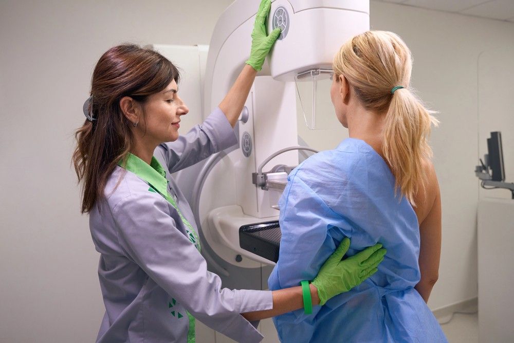
When a woman walks into a breast screening clinic, she often assumes the mammogram sees everything in her breast. But here’s a common question: do 3D mammograms show lymph nodes? It’s a smart question. Lymph nodes play a critical role in breast health and cancer staging.
In this article, we’ll peel back the layers (pun intended) and explain what 3D mammography can — and cannot — do when it comes to lymph nodes. We’ll also highlight how NextGen Diagnostic Imaging near Mid West handles this in real clinical practice.
Understanding Mammography Basics
Mammography is the workhorse of breast screening. Traditional 2D mammograms capture a flat image of the breast in two views (craniocaudal and mediolateral). But overlapping tissue can mask lesions.
Enter 3D mammography, also known as digital breast tomosynthesis: this technology acquires multiple low-dose images from different angles, which are then reconstructed into a pseudo-3D stack of thin slices. That reduces overlap and improves detection of small lesions.

What Are Lymph Nodes and Why They Matter
To understand whether 3D mammograms can show lymph nodes, first recall what lymph nodes are. The lymphatic system is like the body’s drainage network — it carries lymph fluid, immune cells, and potential cancer cells.
In the breast region, lymph nodes (especially in the axilla, or underarm) are critical pathways for drainage. In breast cancer, cancer cells often travel to these nodes first — so examining them is a key step in staging and treatment.
How Medical Imaging Visualizes Lymph Nodes
So how do radiologists typically visualize lymph nodes?
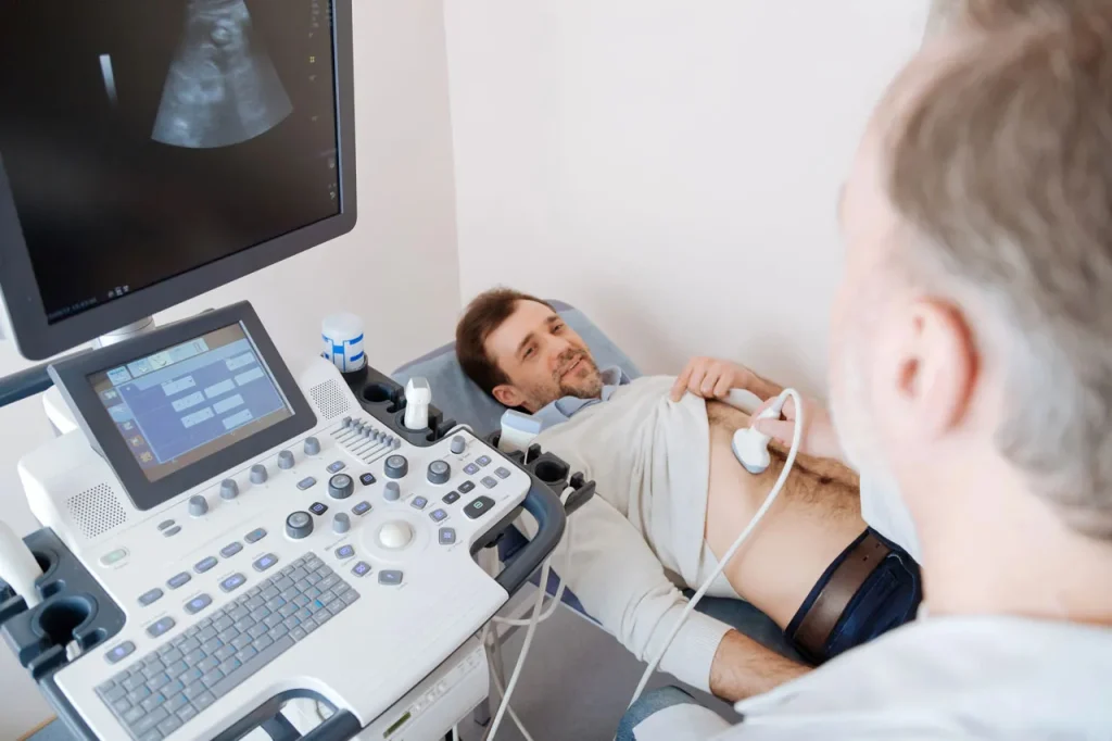
- Ultrasound is often a first go-to: it can distinguish soft tissue and fluid patterns; nodes often appear as oval structures with a fatty hilum.
- MRI provides excellent contrast and depth, especially with contrast agents.
- CT and PET/CT (with radiotracers) are used in oncology staging to catch nodes beyond the breast region.
By contrast, mammograms (2D or 3D) are optimized for breast parenchyma, not deep soft-tissue nodes. Their field of view, imaging geometry, and compression constraints limit node visualization.
Can 3D (Tomosynthesis) Capture Lymph Nodes?
Now to the heart of the matter: can 3D mammography show lymph nodes? The short answer: sometimes, but not reliably or comprehensively.
Physical and technical constraints: When the breast is compressed, much of the underarm region (where many nodes lie) is partially out of the imaging field. Also, the geometry of tomosynthesis is optimized for breast tissue slices, not for extra-breast soft tissues.
Typical field of view: Most mammogram units aim at covering the full breast, including the chest wall as much as possible, but they aren’t designed to fully include the axilla.
Therefore, 3D mammograms might incidentally catch enlarged or superficial nodes near the breast edge, but many nodes — especially deep or posterior ones — won’t be visualized.
What Research Says About 3D Mammograms & Nodes
What does the evidence show?
Some retrospective studies report that mammograms (even 3D) occasionally visualize incidental axillary lymph nodes, particularly when enlarged. However, the sensitivity for detecting pathological nodes is low.
In other words: yes, radiologists sometimes see suspicious node shadows on mammogram slices, but they cannot rely on that to rule in or rule out nodal involvement.
False positives (benign nodes appearing suspicious) and false negatives (diseased nodes not seen) are common.
When Might a 3D Mammogram Show Lymph Nodes?
Under certain conditions, a 3D mammogram may reveal nodes. For example:
- If a node is enlarged, protruding near the breast edge, or pushing into the field of view.
- If it lies in tissue immediately adjacent to the breast, not deeply tucked in the axilla.
- If the imaging protocol or unit has a more generous field capturing some axillary tissue.
Still, these are exceptions, not the rule.
Why You Shouldn’t Rely on 3D Mammograms for Node Detection
Relying on a 3D mammogram to assess lymph nodes is risky.
- Missed deep nodes: Many nodes lie behind muscles, fat, or in regions compressed out of view.
- Radiation trade-offs: Pushing coverage further into the axilla might increase dose or compromise image quality.
- Interpretative challenges: Nodes, fat lobules, and fibroglandular tissues can confuse even seasoned radiologists.
Thus, mammograms are not intended — nor validated — for node staging.
How Radiologists Handle Suspicious Nodes
So what happens when a radiologist thinks they see a suspicious node on a mammogram image (2D or 3D)?
- Flag it in the report as an incidental finding.
- Recommend follow-up imaging, typically an axillary ultrasound. Ultrasound can assess size, shape, cortical thickening, and blood flow.
- If ultrasound is suspicious, proceed with a fine-needle aspiration (FNA) or core biopsy to sample tissue.
Other times, if a patient already has a known breast cancer diagnosis, dedicated breast MRI or PET/CT may be ordered for staging.
Complementary Methods to Detect Lymph Nodes
Because mammograms aren’t enough, clinicians use other imaging methods:
- Breast / axillary ultrasound: Real-time, inexpensive, no radiation — a standard next step.
- MRI: Especially contrast-enhanced MRI can show nodal structures with good contrast.
- PET/CT or sentinel node imaging: Used in oncology for systemic staging, especially beyond the breast region.
These modalities provide better depth, contrast, and field coverage specifically for nodes.
What This Means for You (the Patient)
If you’re getting a 3D mammogram at NextGen Diagnostic Imaging in Mid West, here’s how to think about it:
- Don’t expect the mammogram to evaluate your lymph nodes reliably.
- If your report mentions possible node shadows, ask about axillary ultrasound and further evaluation.
- Understand that mammography is mainly for detecting internal masses, calcifications, and architectural distortions — not node staging.
Having this clarity helps you engage in discussions with radiologists and clinicians.

Why You Should Consider NextGen Diagnostic Imaging in Mid West
Let me tell you why I mention NextGen Diagnostic Imaging. In Mid West, this center offers state-of-the-art 3D tomosynthesis, with expert radiologists trained to spot incidental findings and guide patients properly.
They maintain collaborative protocols: if any suspicious node hint shows up, they seamlessly facilitate ultrasound referrals and biopsies.
Their commitment is to not just screening, but comprehensive breast care, ensuring every patient receives thorough and compassionate evaluation.
How to Prepare for a Mammogram
Let me also drop some tips so your imaging experience is smoother (which helps image quality too):
- Wear a two-piece outfit (top and bottom) so you only remove your top.
- Avoid deodorants, antiperspirants, powders, or creams under the arms or on the breast — they can show up as artifacts.
- Inform the technologist about prior surgeries, implants, or scars in the chest or axilla region.
- Try to schedule your mammogram in the week after your period (when breasts are less tender).
Innovations in Imaging Lymph Nodes
The field is evolving.
- Photon-counting mammography may increase contrast and dynamic range, possibly improving incidental detection.
- Artificial intelligence and deep learning tools are being tested to flag incidental findings, including suspicious nodes.
- Molecular imaging, such as lymph-targeted tracers, may one day allow direct mammographic mapping of sentinel nodes.
These advancements may bridge the gap between breast imaging and nodal evaluation.
Key Takeaways – Do 3D Mammograms Show Lymph Nodes?
Let’s recap:
- 3D mammography is primarily for imaging breast tissue, not lymph nodes.
- Occasionally, enlarged or superficial lymph nodes may be seen, but that’s incidental.
- The sensitivity and specificity for detecting nodal disease via mammogram are limited.
- If a suspicious node is suggested, follow-up imaging (especially ultrasound) and possible biopsy are standard.
- At NextGen Diagnostic Imaging, patients receive the complete continuum of care, from screening to diagnostic evaluation.
NextGen Diagnostic Imaging Serving the Mid West Community and Beyond in Houston
NextGen Diagnostic Imaging is dedicated to serving the diverse needs of the local community of Houston, including individuals residing in neighborhoods like Mid West. With its convenient location near landmarks such as the Briarmeadow Park and major intersections like Beverly Hills St. and Ann Arbor Dr. (coordinates: 29.73019301718039, -95.50934454470142), we offer 3d mammograms Houston services.
Get 3d Mammograms Services At Mid West Now
Navigate from Mid West to NextGen Diagnostic Imaging Now
Conclusion
In short: 3D mammograms are not a reliable tool for assessing lymph nodes. Yes — sometimes a node will peek into the imaging field, but many won’t. Your radiologist won’t, and shouldn’t, use mammography alone to make decisions about nodes.
That’s why secondary imaging exists, and why institutions like NextGen Diagnostic Imaging in Mid West take a holistic approach. If you ever see mention of possible node shadows in your mammogram report, don’t panic — it’s just a clue. Push for an ultrasound or further evaluation, and stay informed. You deserve a full picture of your breast health.
FAQs
1. Can a 3D mammogram replace an ultrasound for checking lymph nodes?
No — a 3D mammogram is not designed to evaluate lymph nodes. Ultrasound remains the standard tool for assessing axillary nodes due to its ability to capture depth, structure, and real-time imaging.
2. If a mammogram image shows something near the underarm, does that mean cancer?
Not necessarily. What appears near the underarm could be fatty tissue, overlapping structures, or benign lymph nodes. Further evaluation, usually via ultrasound or biopsy, is required to determine significance.
3. Will my radiologist always comment on lymph nodes in a mammogram report?
No — because mammograms are not reliable for node assessment, radiologists generally focus their reports on breast findings. If a node-like shadow is seen incidentally, it may be flagged; otherwise, nodes are typically addressed via dedicated imaging.
4. Can MRI detect lymph nodes better than a mammogram?
Yes. MRI, especially contrast-enhanced MRI, offers better soft tissue contrast and a greater field of view, and is more suited for detecting and characterizing lymph nodes when clinically indicated.
5. Are there risks in trying to include lymph node regions in mammogram imaging?
Yes. Extending the imaging field to include more axillary tissue can lead to higher radiation exposure and may degrade image quality in the breast region. It’s not standard practice due to those trade-offs.

NextGen Scans
Be part of a team driven by compassion and a commitment to putting patients first. At NextGen Scans, the care you provide has a meaningful impact every single day.
or Call us at
(346) 487-7037
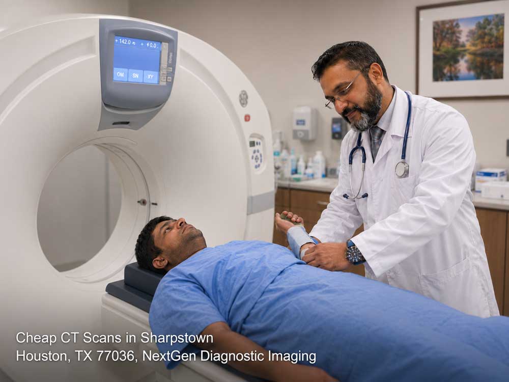
The Symptoms Doctors Almost Missed — Until a CT Scan Changed Everything. Sharpstown
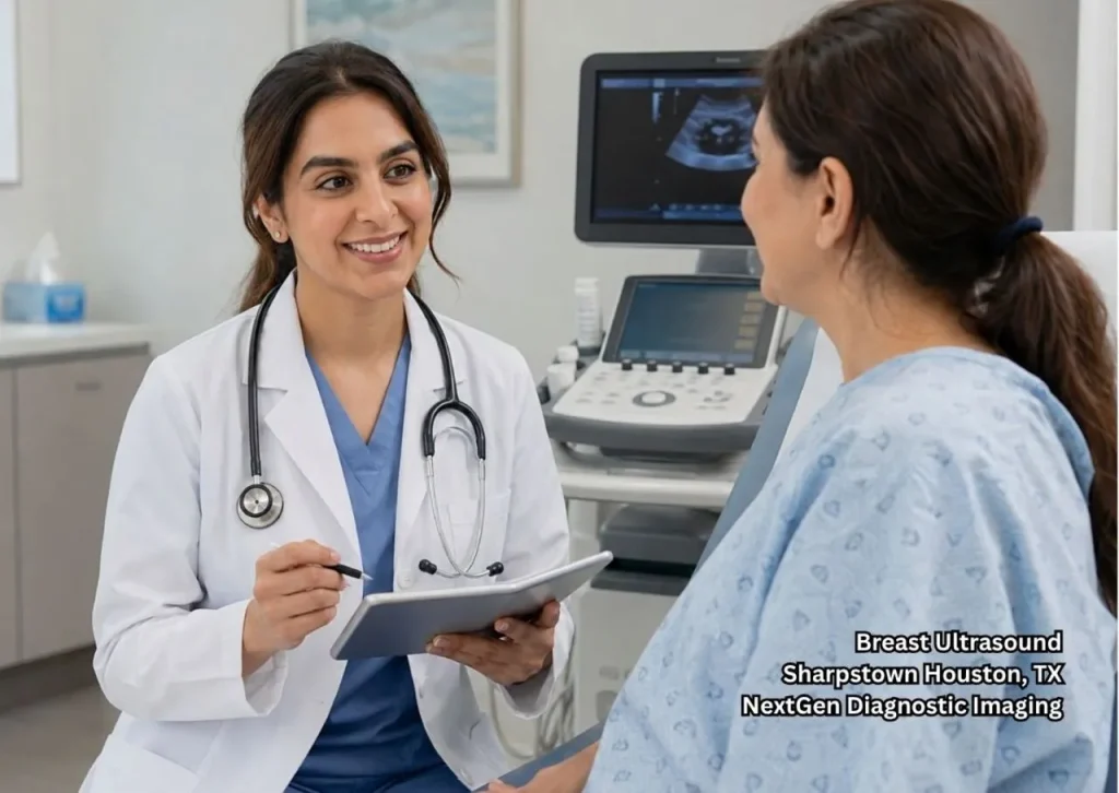
Many symptoms seem minor at first. A headache, stomach pain, dizziness, or ongoing discomfort can easily be linked to everyday life. Yet some symptoms may point to a deeper issue. For many patients, cheap CT scans provide a clearer look inside the body. These scans can reveal conditions that may not appear during a physical…Tired of Waiting and Worrying? A Breast Ultrasound Can Give You Real Answers About That Lump. Sharpstown
Why Finding a Lump Can Feel Scary A breast lump can raise concerns about your health. Many people want answers as soon as possible, and concerns often grow when answers are not available right away. If your doctor recommends a breast ultrasound Houston patients trust, the test can provide helpful information about the lump. Studies…7 Reasons Sharpstown Families Are Finally Choosing Local Imaging Centers Over Big Hospitals in 2026
Introduction Healthcare decisions are changing fast in 2026, especially in Sharpstown. Families are becoming more intentional about where they receive medical imaging services, and many are stepping away from large hospital systems in favor of local imaging centers. Why? Because convenience, affordability, comfort, and personalized care matter more than ever. For years, hospitals were seen…
Contact Us: We’re Here to Help!
We’d love to hear from you! Whether you have questions about our services, need more information on our diagnostic and pain management options, or would like to schedule an appointment, our friendly and professional team is here to assist you every step of the way. We are dedicated to providing you with the best care and support, and we are happy to address any concerns or inquiries you may have.