Is Ultrasound Better For Dense Breasts?
June 23, 2025
If you’ve been told you have dense breasts, you’re probably wondering: “Does that mean something’s wrong? Should I get an ultrasound instead of a mammogram?” You’re not alone—and the short answer is: ultrasound can often be a powerful supplemental tool for women with dense breast tissue.
In this article, we’ll break everything down in plain English and help you understand why centers like NextGen Diagnostic Imaging often recommend ultrasound for clearer results.
Understanding Breast Density
What Are Dense Breasts?
Breast density has nothing to do with how your breasts feel. It refers to how your breast tissue appears on a mammogram. If you have more glandular and fibrous tissue compared to fatty tissue, your breasts are considered dense.
How Common Is Breast Density?
Very. In fact, nearly 50% of women over the age of 40 have dense breasts. It’s completely normal, but it can complicate routine screenings.
Why Breast Density Matters in Screening
The Challenge with Mammograms
Here’s the thing: mammograms are great, but dense tissue shows up white on a mammogram—and guess what else shows up white? Cancer. That’s like trying to find a snowflake in a snowstorm. Not ideal.
Risk Factors Tied to Dense Breast Tissue
Having dense breasts doesn’t just make detection tricky—it also slightly increases your risk for developing breast cancer. That’s why better screening methods, like ultrasound, are gaining attention.
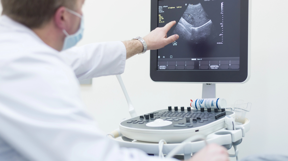
What Is an Ultrasound?
How It Works
Ultrasound uses sound waves (not radiation) to create detailed images of your breast tissue. A technician glides a small probe over your skin, and voila—real-time visuals of what’s beneath the surface.
Types of Breast Ultrasound
- Handheld ultrasound – The traditional method, used by a trained technician.
- Automated Breast Ultrasound (ABUS) – A high-tech version offering full breast imaging in a consistent way.
Ultrasound vs Mammogram for Dense Breasts
When it comes to screening women with dense breasts, the conversation often turns to mammograms versus ultrasound. Each method has its strengths and limitations, and understanding these can help you and your doctor make better screening decisions—especially if you have dense breast tissue.
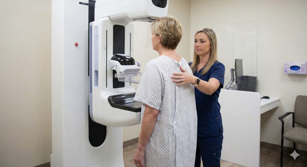
Pros and Cons of Mammograms
Pros:
- Standard screening tool:
Mammograms are the gold standard for breast cancer screening. Most guidelines still recommend starting annual or biennial mammograms at age 40 (or earlier for high-risk individuals). They’ve saved countless lives by detecting cancers early—when they’re most treatable. - Good at spotting calcifications (early signs of cancer):
One of the unique strengths of mammography is its ability to identify microcalcifications—tiny deposits of calcium in breast tissue. These can be early indicators of ductal carcinoma in situ (DCIS), a type of pre-cancer that ultrasound might miss entirely.
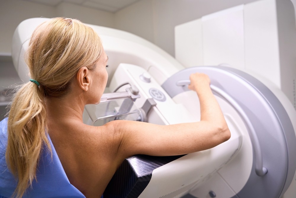
Cons:
- Less effective in dense tissue:
Dense breast tissue appears white on a mammogram—just like tumors. This lack of contrast makes it difficult to distinguish between normal dense tissue and potential cancer, especially in early stages. - Radiation exposure (though minimal):
While the radiation dose from a mammogram is very low, it’s still a factor to consider, especially for women needing frequent screenings or those with a strong family history of breast cancer. - Can miss some cancers hiding in dense areas:
Unfortunately, because of the overlap between dense tissue and tumors on the scan, cancers can be completely hidden on a mammogram. This is why supplemental imaging is so important for women with dense breasts.
Pros and Cons of Ultrasound
Pros:
- No radiation involved:
One of the biggest benefits of ultrasound is that it uses sound waves, not radiation. This makes it an excellent option for younger women, women who are pregnant, or those seeking to limit their cumulative radiation exposure. - Excellent at spotting tumors that mammograms miss:
Ultrasound provides a different view of breast tissue. It’s especially effective in visualizing masses in dense tissue, which appear more clearly due to the contrast between solid and fluid-filled areas. This makes ultrasound a valuable follow-up tool after a mammogram. - Great for younger women with naturally denser tissue:
Women under 40 often have higher breast density, making mammograms less effective. Ultrasound offers a safer and more revealing screening option for this age group, especially when there are symptoms like lumps or pain.
Cons:
- Higher chance of false positives:
Ultrasound can be very sensitive, but that’s a double-edged sword. It may identify non-cancerous lumps that require further evaluation—leading to extra appointments, stress, or even unnecessary biopsies. - Not meant to replace mammograms entirely:
Ultrasound should be seen as a complement to mammograms, not a substitute. It’s particularly useful when mammography results are unclear or when screening women with dense breast tissue, but it doesn’t provide a full picture on its own. - May require additional tests to confirm findings:
Because ultrasound isn’t as precise in categorizing abnormalities, any suspicious area might need follow-up with MRI, biopsy, or a more detailed mammogram. This can increase both cost and patient anxiety, even when results are benign.
Sensitivity in Dense Tissue
Ultrasound excels at detecting tumors in dense breasts, where mammograms may fall short. In fact, multiple studies have found that adding ultrasound to mammography increases cancer detection rates significantly. According to research, it can reveal 2–3 additional cancers per 1,000 women that mammograms alone would miss. That may not sound like a lot, but when early detection saves lives, every single case matters.
Ultrasound can differentiate between solid masses and cysts, providing clarity when mammograms offer nothing but a white blur. This makes it particularly effective in catching invasive cancers that might not calcify or be large enough to show on a mammogram.
False Positives and Overdiagnosis
Here’s where things get a bit murky. Ultrasound can over-detect, meaning it finds things that aren’t cancer but still look suspicious enough to require further testing. This leads to:
- Unnecessary biopsies
- Extra imaging appointments
- Increased anxiety
This is called a false positive, and while it doesn’t harm your body, it can take a toll mentally and financially. There’s also a slight risk of overdiagnosis, where something benign is treated aggressively just in case.
That said, many women say they’d rather go through extra tests than risk missing something important. For patients with a family history of breast cancer or previous abnormal results, the peace of mind alone can make the false positives worth it.
When Ultrasound Becomes a Smart Addition
Supplemental Screening Explained
If you’ve had a “normal” mammogram but still have dense breasts, ultrasound is a logical next step. It doesn’t replace mammograms—it complements them.
Guidelines from Medical Experts
Organizations like the American College of Radiology and the Society of Breast Imaging support personalized screening strategies, especially for women with dense breasts.
Why NextGen Diagnostic Imaging Recommends Ultrasound
Patient-Centered Approach
At NextGen Diagnostic Imaging, they don’t just scan and send you on your way. Their team tailors each screening to your personal risk profile and tissue type.
Advanced Technology and Care
They use state-of-the-art ultrasound machines and employ experienced technicians trained specifically for breast imaging. You’re not just another chart—you’re a person they care about getting answers for.
Real Stories, Real Results
Case Study: Maria’s Experience
Maria, age 42, had a clear mammogram—but persistent pain in one breast. Her OB-GYN referred her to NextGen Diagnostic Imaging for an ultrasound. What they found? A small, early-stage tumor that didn’t show up on her mammogram.
Today, Maria is cancer-free—thanks to that extra layer of screening.
What Patients Say
Many women who’ve used NextGen’s services praise the comfort, clarity, and professionalism of the entire experience. One patient said, “It felt like they cared about finding what others missed.”
How to Know If You Have Dense Breasts
Reading Your Mammogram Report
After your mammogram, your report will usually mention your breast density. Look for terms like:
- “Heterogeneously dense”
- “Extremely dense”
These suggest your breasts could benefit from supplemental imaging like ultrasound.
Talking with Your Doctor
Don’t hesitate to ask: “Should I consider an ultrasound based on my breast density?” The more proactive you are, the better your chances of early detection.
Are There Alternatives to Ultrasound?
MRI
Magnetic Resonance Imaging (MRI) is another option, especially for high-risk women. It’s extremely sensitive but also more expensive and time-consuming.
3D Mammography (Tomosynthesis)
A step up from standard mammograms, 3D mammography takes layered images for a clearer picture. Still, even this tech can miss things in very dense tissue—so many women opt for both 3D and ultrasound.
Insurance and Cost Considerations
Is Ultrasound Covered by Insurance?
It depends on your location and your provider. In many states, laws require insurance to cover ultrasound if you have dense breasts. It’s worth checking your policy or asking the staff at NextGen Diagnostic Imaging—they’ll help you sort it out.
Cost Without Coverage
Out-of-pocket, a breast ultrasound can range from $100 to $400. It’s a small price to pay for peace of mind and potentially life-saving detection.
NextGen Diagnostic Imaging Serving the Gulfton Community and Beyond in Houston
NextGen Diagnostic Imaging is dedicated to serving the diverse needs of the local community of Houston, including individuals residing in neighborhoods like Gulfton. With its convenient location near landmarks such as the Benavidez Elementary School and major intersections like Rampart St. & Gulfton St. (coordinates: 29.716703999287475, -95.48863238108788), we offer breast ultrasound Houston services.
Get Breast Ultrasound Houston Services at Gulfton Now
Navigate from Gulfton to NextGen Diagnostic Imaging Now
The Clearer Path to Confident Breast Health Decisions
Is ultrasound better for dense breasts? Yes—especially as a backup to your routine mammogram. It’s not about picking one over the other—it’s about creating a screening combo that works for your body. If you have dense tissue, you deserve a clearer picture—and ultrasound can give you that.
At NextGen Diagnostic Imaging, it’s not just about machines and images—it’s about finding answers, early detection, and peace of mind. So if you’re navigating your next screening, ask if ultrasound is right for you. Chances are, it just might be the tool that makes the difference.
FAQs
1. Can I skip my mammogram if I get an ultrasound instead?
Nope. Ultrasound is a supplement—not a substitute. You still need regular mammograms.
2. How do I know if I have dense breasts?
Your mammogram report will tell you. Look for terms like “heterogeneously dense” or “extremely dense.”
3. Is ultrasound painful or uncomfortable?
Not at all. It’s non-invasive, radiation-free, and generally pain-free.
4. Are all imaging centers qualified for breast ultrasound?
Not necessarily. Always choose a reputable center like NextGen Diagnostic Imaging with breast imaging expertise.
5. How often should I get a breast ultrasound?
This depends on your risk level and what your doctor recommends. Some women get it yearly as a supplement.

NextGen Scan
How X-Ray Services Help Doctors Make Fast Diagnoses
In healthcare, timing can be the difference between a minor concern and a major medical emergency. When symptoms appear—whether it’s sudden pain, shortness of breath, or an unexplained injury—doctors need answers quickly. This is where X-ray services step in as one of the most powerful diagnostic tools in modern medicine. For patients seeking X-ray services…What Are Mammogram Services And What Do They Include?
When it comes to protecting your long-term health, few screenings are as powerful—or as misunderstood—as a mammogram. Mammogram services are not just routine medical appointments; they are proactive tools that help detect breast changes long before symptoms appear. For women in Houston and Shenandoah, access to high-quality mammogram services can mean the difference between early…What Makes an Ultrasound Affordable Without Sacrificing Accuracy?
When patients hear that an ultrasound is “affordable,” the word often triggers doubt. In a healthcare system where prices vary wildly, many people assume lower cost must mean lower quality. After all, we’ve been trained to believe that better care always costs more. But diagnostic imaging doesn’t work that way. Ultrasound accuracy isn’t determined by…


Contact Us: We’re Here to Help!
We’d love to hear from you! Whether you have questions about our services, need more information on our diagnostic and pain management options, or would like to schedule an appointment, our friendly and professional team is here to assist you every step of the way. We are dedicated to providing you with the best care and support, and we are happy to address any concerns or inquiries you may have.