What Does a Chest X-ray Show? Understanding Its Role in Diagnosis
April 26, 2025
A chest X-ray (CXR) is one of the most common and effective diagnostic tools used by healthcare professionals to examine the internal structures of the chest. This includes the heart, lungs, blood vessels, ribs, and the area around the chest. X-rays work by passing a small amount of radiation through the body, capturing images of these structures on film or digital receptors. A chest X-ray is typically ordered when a patient presents symptoms like chest pain, persistent cough, shortness of breath, or unexplained weight loss. It can also be used as a routine part of a physical exam or to monitor the progression of known health conditions.
In this blog post, we will explore in detail what a chest X-ray shows, how it works, and how it aids in diagnosing a variety of medical conditions.
What Can a Chest X-ray Reveal?
A chest X-ray provides a detailed image of the chest area, allowing doctors to assess various organs and structures within the chest cavity. Here’s a closer look at the main components that a chest X-ray can reveal:
1. The Lungs
The most common reason for a chest X-ray is to examine the lungs. This imaging test can help detect several lung-related conditions, including:
- Pneumonia: Infections such as pneumonia cause inflammation in the lungs, which can be identified through abnormalities in the lung tissue.
- Tuberculosis (TB): TB can lead to cavities or scar tissue in the lungs, which is visible on X-ray images.
- Lung Cancer: Tumors or nodules in the lungs may be detected through chest X-rays, although further tests are usually required to confirm the presence of cancer.
- Chronic Obstructive Pulmonary Disease (COPD): Chest X-rays can reveal emphysema or signs of lung damage caused by smoking and other environmental factors.
2. The Heart
While chest X-rays primarily focus on the lungs, they also provide valuable information about the heart. A chest X-ray can show:
- Heart Enlargement: An enlarged heart, often a sign of heart disease or heart failure, can be detected by changes in the size and shape of the heart.
- Fluid Buildup: Fluid in the lungs or around the heart, often a sign of heart failure, can be observed on X-ray images.
- Aortic Aneurysm: A dilated aorta, which may indicate an aneurysm, can also be seen on a chest X-ray.
3. The Ribs and Chest Wall
Chest X-rays provide detailed views of the ribs and chest wall. Doctors can use this information to:
- Detect Fractures: Broken or fractured ribs, which might not be immediately obvious, are visible on an X-ray.
- Assess Other Injuries: Other chest wall injuries, including damage to muscles and tissues, can be detected through a chest X-ray.
4. The Diaphragm
The diaphragm is a large muscle that separates the chest and abdominal cavities. Changes in the shape or position of the diaphragm can indicate certain health conditions, including:
- Hernia: A hernia can cause abnormal positioning of the diaphragm, which may be visible on an X-ray.
- Liver or Stomach Disorders: Abnormalities in the diaphragm may also suggest issues with the liver, stomach, or other organs in the abdominal cavity.
Why a Chest X-ray is Done: Common Reasons for a Chest X-ray
Chest X-rays are used for a variety of diagnostic purposes, from checking for conditions related to lung function to assessing trauma-related injuries. Below are some common reasons a healthcare provider may recommend a chest X-ray:
1. Diagnosing Respiratory Conditions
Doctors often use chest X-rays to investigate symptoms such as persistent cough, chest pain, or difficulty breathing. Common respiratory conditions that a chest X-ray can detect include:
- Pneumonia: Inflammation in the lungs caused by infection.
- Pulmonary Edema: Fluid buildup in the lungs, often due to heart failure.
- Lung Cancer: Tumors in the lungs or other lung-related abnormalities.
2. Monitoring Heart Health
A chest X-ray can be an essential tool in evaluating heart health, especially for individuals with symptoms of heart disease, such as chest pain or difficulty breathing. It helps assess conditions like:
- Heart Enlargement: As a result of heart disease or heart failure.
- Fluid Accumulation: A sign of heart failure or severe lung disease.
3. Detecting Broken Ribs or Other Injuries
If a patient has experienced trauma to the chest, such as in a car accident or fall, a chest X-ray can quickly identify broken or fractured ribs. This helps doctors determine the extent of injury and appropriate treatment.
4. Evaluating Treatment Progress
For individuals undergoing treatment for certain lung or heart conditions, doctors may order chest X-rays to track progress and monitor any changes. This could include conditions like lung cancer or heart failure.
How a Chest X-ray Works: The Process and Technology
A chest X-ray works by using radiation to create images of the chest area. The X-ray machine sends a controlled amount of radiation through the body, which passes through the chest and is absorbed by different tissues in varying amounts. These differences are captured by a detector on the other side of the body, which converts the data into an image.
The Procedure: Step-by-Step
- Preparation: You may be asked to remove jewelry or clothing that could interfere with the image.
- Positioning: You will be asked to stand in front of the X-ray machine, often in a position where your chest faces the machine and your side is also captured.
- Imaging: You will be asked to hold your breath briefly while the X-ray is taken. This helps produce clearer images.
- Post-Procedure: The images are analyzed by a radiologist, and results are shared with your healthcare provider for further evaluation.
Interpreting Chest X-ray Results
Once the chest X-ray is taken, the images are sent to a radiologist who specializes in interpreting X-rays. The radiologist will look for abnormal patterns or changes in the chest area that may indicate the presence of a condition. Some of the key factors considered during interpretation include:
- Size and Shape of the Heart: Enlargement may suggest heart disease.
- Lung Tissue Density: Areas of increased or decreased density can indicate infections, cancer, or fluid buildup.
- Bone Structure: Fractures or abnormalities in the ribs or spine are carefully examined.
- Diaphragm and Surrounding Tissues: Changes in diaphragm shape or position can signal underlying issues.
What Chest X-rays Can’t Show
While chest X-rays are a powerful diagnostic tool, they have limitations. There are certain conditions and details that may not be visible on a chest X-ray, including:
- Early-Stage Lung Cancer: Small or early tumors might not be detectable in an X-ray.
- Heart Valve Problems: X-rays can show heart enlargement but are less effective in detecting specific heart valve issues.
- Soft Tissue Damage: Conditions affecting soft tissues, such as muscle strains, may not be visible.
In such cases, additional imaging tests like CT scans, MRIs, or ultrasounds may be required for a more detailed diagnosis.
Nextgen Diagnostics Serving the Gulfton Community and Beyond in Houston
Nextgen Diagnostics is dedicated to serving the diverse needs of the local community of Houston, including individuals residing in neighborhoods like Gulfton. With its convenient location near landmarks such as the Rodriguez Elementary Spark Park and major intersections like Gulfton St & Chimney Rock Rd (coordinates: 29.716766785154757, -95.47829750765696), we offer Cheap CT Scans Houston services.
Get Cheap CT Scans Houston Services at Gulfton Now
Navigate from Gulfton to Nextgen Diagnostics Now
The Importance of Chest X-rays in Diagnosing Health Conditions
Chest X-rays play a significant role in diagnosing a wide range of health conditions, especially those affecting the lungs, heart, and chest wall. They are quick, non-invasive, and provide valuable insights into a patient’s health status. Whether it’s diagnosing pneumonia, assessing heart function, or detecting fractures, chest X-rays are essential in helping healthcare providers make informed decisions about patient care.
If you need a chest X-ray, Nextgen Diagnostics offers reliable and accurate imaging services to help you understand your health better and get the treatment you need.
Frequently Asked Questions
- What conditions can a chest X-ray detect? A chest X-ray can detect a variety of conditions, including pneumonia, tuberculosis, lung cancer, broken ribs, heart enlargement, and fluid buildup around the heart. It is also useful for monitoring chronic conditions such as COPD.
- Is a chest X-ray painful? No, a chest X-ray is a non-invasive and painless procedure. The only discomfort might come from having to hold your breath for a few seconds during the imaging process.
- How long does a chest X-ray take? A chest X-ray typically takes only a few minutes. The actual imaging process is quick, and you may be asked to wait briefly for the radiologist to review the images.
- Are there risks associated with chest X-rays? While chest X-rays use radiation, the amount is very small and generally considered safe for most people. However, pregnant women and young children may need special consideration or alternative imaging methods.
- How do I prepare for a chest X-ray? No special preparation is needed for a chest X-ray. You may be asked to remove jewelry or clothing that could interfere with the imaging, and you’ll be asked to stand in front of the X-ray machine during the procedure.

NextGen Scan
How X-Ray Services Help Doctors Make Fast Diagnoses
In healthcare, timing can be the difference between a minor concern and a major medical emergency. When symptoms appear—whether it’s sudden pain, shortness of breath, or an unexplained injury—doctors need answers quickly. This is where X-ray services step in as one of the most powerful diagnostic tools in modern medicine. For patients seeking X-ray services…What Are Mammogram Services And What Do They Include?
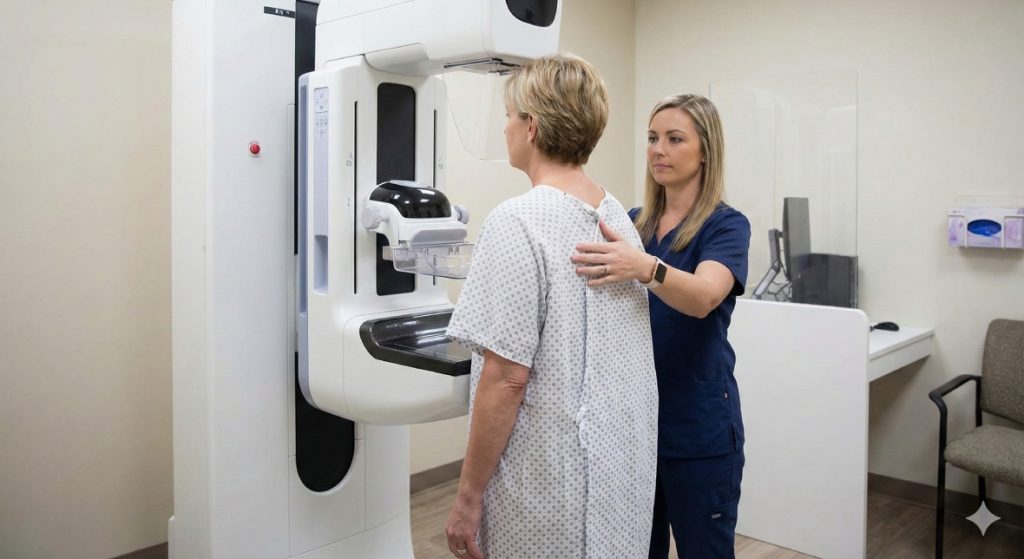
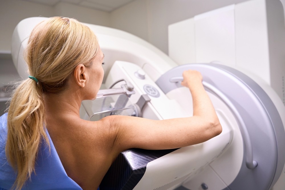
When it comes to protecting your long-term health, few screenings are as powerful—or as misunderstood—as a mammogram. Mammogram services are not just routine medical appointments; they are proactive tools that help detect breast changes long before symptoms appear. For women in Houston and Shenandoah, access to high-quality mammogram services can mean the difference between early…What Makes an Ultrasound Affordable Without Sacrificing Accuracy?
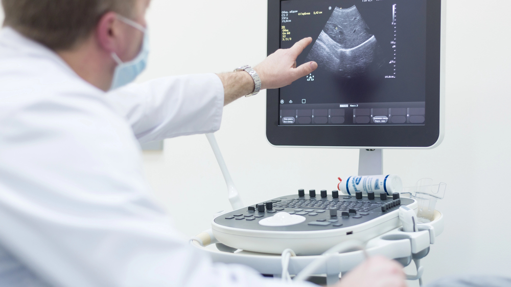
When patients hear that an ultrasound is “affordable,” the word often triggers doubt. In a healthcare system where prices vary wildly, many people assume lower cost must mean lower quality. After all, we’ve been trained to believe that better care always costs more. But diagnostic imaging doesn’t work that way. Ultrasound accuracy isn’t determined by…


Contact Us: We’re Here to Help!
We’d love to hear from you! Whether you have questions about our services, need more information on our diagnostic and pain management options, or would like to schedule an appointment, our friendly and professional team is here to assist you every step of the way. We are dedicated to providing you with the best care and support, and we are happy to address any concerns or inquiries you may have.