When Were Ultrasounds Invented?
June 24, 2025
A Quick Peek into Medical Imaging History
Before we dive into the fascinating journey of ultrasound invention, let’s zoom out for a second. Picture this: a world where doctors couldn’t see inside the body without making a cut. That was reality for centuries. But then came X-rays, CT scans, MRIs, and yes—ultrasounds. Each of these tools rewrote the medical playbook. But one stands out for being safe, radiation-free, and incredibly versatile: the ultrasound.
What Exactly Is an Ultrasound?
Understanding the Basics
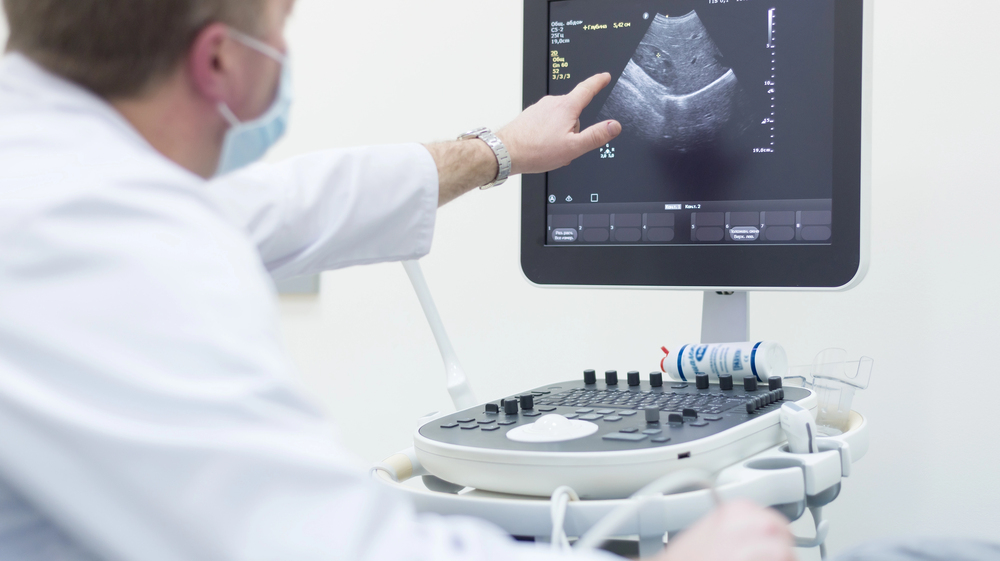
Ultrasound is a form of imaging that uses high-frequency sound waves to produce images of the inside of the body. Think of it like sonar or echolocation—similar to how bats navigate. These waves bounce off tissues and return to the transducer, which converts them into real-time visuals. Pretty amazing, right?
How Ultrasounds Work Today
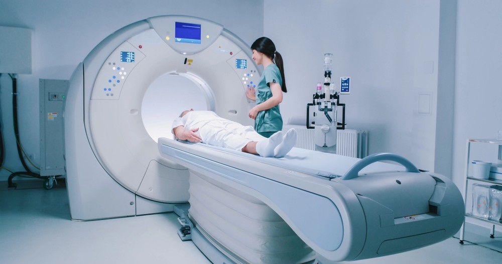
Modern ultrasound machines are sleek, powerful, and even portable. From checking fetal growth to spotting tumors and guiding needle biopsies, ultrasound has become a diagnostic superhero—quiet, safe, and precise.
When Were Ultrasounds Actually Invented?
The Origins: Echoes from the Early 1900s
Believe it or not, the roots of ultrasound trace back to the early 20th century, grounded in a powerful scientific principle called the piezoelectric effect. This effect—discovered in 1880 by French physicists Pierre and Jacques Curie—explains how certain crystals produce an electric charge when compressed. Fast-forward a few decades, and this discovery became the foundation for technologies that could send and receive sound waves.
Initially, the piezoelectric effect was used in sonar and industrial applications. Medical use wasn’t even on the radar—yet. But as engineers and scientists began realizing the potential of high-frequency sound waves to interact with solid materials, it planted the seeds for what would eventually become medical ultrasound. These early ideas began to take shape in Europe and Japan, where physicists experimented with sound wave transmission and reflection through different materials—including biological tissue.
So while the actual medical ultrasound machine wasn’t invented in the early 1900s, the theoretical framework and physical tools for ultrasound imaging were already being explored. It was only a matter of time before someone asked: “Can we use this inside the human body?”
The 1940s: WWII and the Spark of Medical Use
World War II didn’t just reshape global politics—it also accelerated technological innovation like never before. One of the most important wartime advancements? Sonar, or Sound Navigation and Ranging. Used to detect enemy submarines beneath the ocean surface, sonar relied on sending sound waves through water and interpreting their echoes.
After the war, brilliant minds began connecting the dots. If sound could reveal the location and shape of submarines, couldn’t it also help visualize structures inside the human body? Enter the era of post-war experimentation, where physicists, engineers, and doctors began repurposing sonar principles for medical imaging.
By the late 1940s, pioneers like Karl Dussik in Austria began publishing research on using ultrasound to visualize the human brain. Although his images were crude and inconsistent, they marked the first documented attempts to apply ultrasound for medical diagnosis. Around the same time, scientists in Japan and the U.S. were conducting parallel research—each team refining the technology in different ways.
This was a turning point in medical history. What had once been a military tool was being reimagined as a life-saving diagnostic method.
1950s–1960s: The First Clinical Applications
By the 1950s, ultrasound technology took a giant leap from the lab bench to the hospital floor. Machines were still massive and far from user-friendly, but they were starting to prove their value in clinical settings.
One of the earliest and most impactful applications? Obstetrics. Doctors began using ultrasound to monitor pregnancies. This was revolutionary. Until then, fetal development could only be guessed through manual palpation, external measurements, or X-rays—which exposed both mother and fetus to radiation.
Enter Dr. Ian Donald, a Scottish obstetrician widely credited with developing the first practical ultrasound machine for clinical use. In 1958, he and engineer Tom Brown published a groundbreaking paper describing how ultrasound could differentiate between various abdominal masses. This led to the creation of a prototype scanner that could produce real-time images of a fetus—a moment that changed prenatal care forever.
Throughout the 1960s, ultrasound systems evolved rapidly. They became more portable, produced clearer images, and were adopted by more medical departments. Cardiology, hepatology, and vascular medicine began using ultrasound for non-invasive diagnosis, adding more momentum to its growth.
By the end of the 1960s, ultrasound had gone from a curiosity to a core diagnostic tool—all within two decades. What started with echoes and war tech had now found a peaceful, powerful purpose in healing and human care.
Who Invented the Ultrasound?
Key Pioneers in Ultrasound History
Ultrasound wasn’t born from just one mind. It was a global evolution. In Austria, Karl Dussik began experimenting with ultrasonic waves for brain imaging in the 1940s. Meanwhile, engineers in Japan, Sweden, and the U.S. were refining the tech.
Dr. Ian Donald and Obstetric Ultrasound
But if we had to crown one figure as the father of modern ultrasound, it’d be Dr. Ian Donald from Scotland. In the late 1950s, he used ultrasound to examine abdominal masses and, soon after, pregnancies. His work laid the groundwork for the field of obstetric ultrasound—something every parent today is familiar with.
How Ultrasound Technology Evolved Over Time
From A-Mode to B-Mode Scanning
Early ultrasounds were A-mode (amplitude mode), which produced one-dimensional images. Then came B-mode (brightness mode), which gave us 2D visuals. It was like going from Morse code to television overnight.
Color Doppler and 3D Imaging
The 1980s and 1990s saw the rise of Doppler ultrasound, which could show blood flow in real time using color-coded visuals. It became a game-changer for cardiologists and vascular specialists.
The Rise of 4D Ultrasound and Real-Time Imaging
And then came 3D and 4D ultrasounds. These weren’t just cool to look at—they gave doctors richer, more accurate insights into the body’s function, all in real time.
Modern-Day Ultrasounds: A Diagnostic Powerhouse
Applications Beyond Pregnancy
While most people think of baby scans, ultrasound is used across dozens of medical fields. It checks organs, joints, blood vessels, muscles, and even guides surgical tools. It’s a jack-of-all-trades—and a master, too.
Portable and Point-of-Care Devices
With today’s tech, we now have handheld ultrasounds. That means doctors can bring imaging to the patient’s bedside—or even into remote villages around the world.
Why Ultrasounds Are a Game-Changer
Non-Invasive and Radiation-Free
Ultrasound doesn’t use ionizing radiation, which means it’s safer, especially for developing babies and repeated use. It’s like having X-ray vision—minus the risks.
Cost-Effective and Safe
Compared to MRI and CT scans, ultrasounds are budget-friendly. That’s why they’re used in small clinics, emergency rooms, and major hospitals alike. And they’re so safe, they’re even used in physical therapy.
The Role of NextGen Diagnostic Imaging Today
Leading the Way with Advanced Technology
At NextGen Diagnostic Imaging, we carry this legacy of innovation forward. Our ultrasound technology combines precision with comfort, ensuring that every scan is as quick and accurate as possible. Whether it’s a routine pregnancy check or complex vascular mapping, we’re ready.
Patient-Centered Care from the Ground Up
What sets NextGen apart isn’t just the tools we use—but how we use them. Our team of specialists prioritizes patient experience, translating complex images into clear answers. We blend tech with trust, every step of the way.
Looking Ahead: What’s the Future of Ultrasound?
AI-Enhanced Imaging
Artificial intelligence is already being integrated into ultrasound machines. Algorithms can now spot patterns in real time, helping radiologists detect issues faster and with greater confidence.
Handheld Devices and Global Access
Portable ultrasound is democratizing healthcare. Soon, even a smartphone could host diagnostic-quality imaging, transforming care in rural and underserved areas. The future? It’s already knocking.
NextGen Diagnostic Imaging Serving the Sharpstown Community and Beyond in Houston
NextGen Diagnostic Imaging is dedicated to serving the diverse needs of the local community of Houston, including individuals residing in neighborhoods like Sharpstown. With its convenient location near landmarks such as the The College of Health Care Professions and major intersections like Southwest Fwy. & Bellaire Blvd. (coordinates: 29.703212665392144, -95.51451719327261), we offer breast ultrasound Houston services.
Get Breast Ultrasound Houston Services at Sharpstown Now
Navigate from Sharpstown to NextGen Diagnostic Imaging Now
The Echo of Innovation
When were ultrasounds invented? Technically, the journey began over a century ago—but it’s still unfolding today. What started as sonar-inspired curiosity has evolved into one of the most essential tools in modern medicine. With each advancement, ultrasounds become faster, clearer, safer, and more powerful.
And at NextGen Diagnostic Imaging, we’re proud to be part of that journey—helping patients every day through cutting-edge diagnostic care with a personal touch. Ultrasound may have been invented decades ago, but its best days are still ahead.
FAQs
1. Who was the first person to use ultrasound in medicine?
Karl Dussik is often credited as the first to use ultrasound medically, particularly for brain imaging in the 1940s.
2. When did ultrasound start being used for pregnancy?
Ultrasound started being used for pregnancy in the late 1950s, largely due to the work of Dr. Ian Donald in Scotland.
3. Are modern ultrasounds different from those in the 1960s?
Absolutely. Today’s ultrasounds offer 3D/4D imaging, Doppler analysis, and AI integration—light years ahead of the basic 2D scans of the 1960s.
4. Can ultrasounds detect cancer?
Yes, they can help detect and monitor tumors, especially in organs like the liver, kidneys, and thyroid, though other imaging or biopsies may be needed for confirmation.
5. Does NextGen Diagnostic Imaging offer Doppler ultrasound?
Yes! At NextGen Diagnostic Imaging, we offer a full range of ultrasound services—including Doppler imaging for vascular and cardiac evaluations.

NextGen Scan
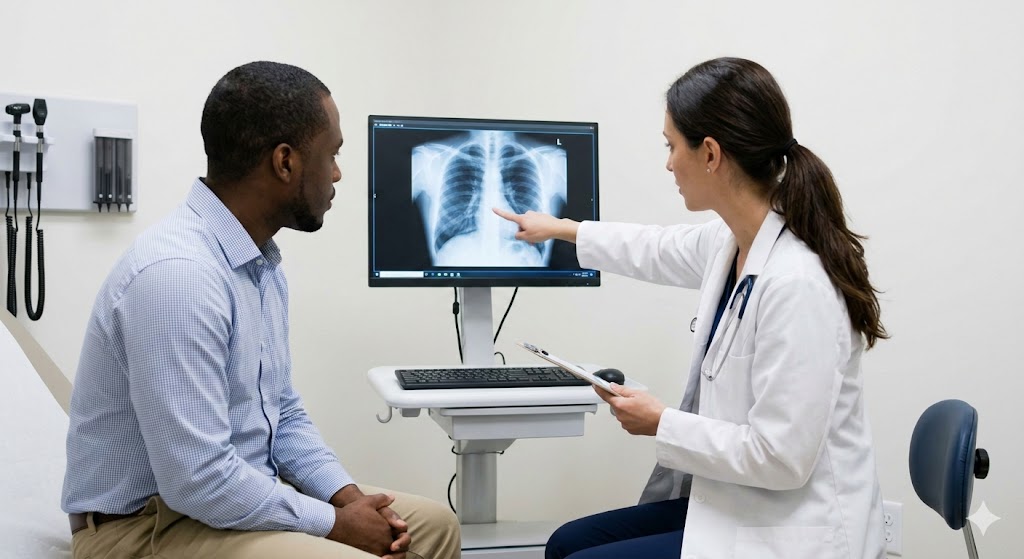
How X-Ray Services Help Doctors Make Fast Diagnoses
In healthcare, timing can be the difference between a minor concern and a major medical emergency. When symptoms appear—whether it’s sudden pain, shortness of breath, or an unexplained injury—doctors need answers quickly. This is where X-ray services step in as one of the most powerful diagnostic tools in modern medicine. For patients seeking X-ray services…What Are Mammogram Services And What Do They Include?
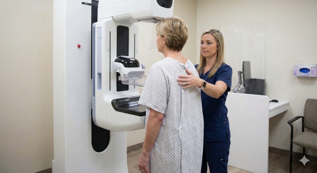
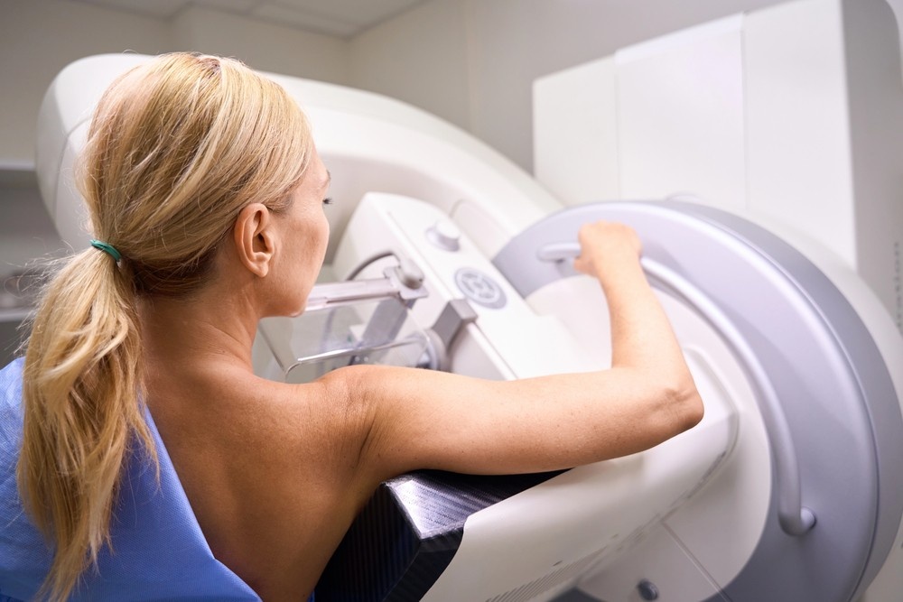
When it comes to protecting your long-term health, few screenings are as powerful—or as misunderstood—as a mammogram. Mammogram services are not just routine medical appointments; they are proactive tools that help detect breast changes long before symptoms appear. For women in Houston and Shenandoah, access to high-quality mammogram services can mean the difference between early…What Makes an Ultrasound Affordable Without Sacrificing Accuracy?
When patients hear that an ultrasound is “affordable,” the word often triggers doubt. In a healthcare system where prices vary wildly, many people assume lower cost must mean lower quality. After all, we’ve been trained to believe that better care always costs more. But diagnostic imaging doesn’t work that way. Ultrasound accuracy isn’t determined by…


Contact Us: We’re Here to Help!
We’d love to hear from you! Whether you have questions about our services, need more information on our diagnostic and pain management options, or would like to schedule an appointment, our friendly and professional team is here to assist you every step of the way. We are dedicated to providing you with the best care and support, and we are happy to address any concerns or inquiries you may have.